In the rapidly evolving world of biotechnology, few discoveries have had as profound an impact as the Green Fluorescent Protein (GFP). Originally discovered in the jellyfish Aequorea victoria, GFP has become an indispensable tool in molecular biology, genetic engineering, and live-cell imaging.
But why is GFP so important in science and research today? And how has it transformed the way we study cells and genes?
Let’s explore how GFP biotechnology is helping researchers see life in color literally.
What Makes GFP So Special?
GFP is a naturally fluorescent protein that emits bright green light when exposed to blue or UV light. This unique feature allows scientists to track and visualize cellular events in real time, without the need for toxic dyes or stains.
Thanks to genetic engineering, the GFP gene can be inserted into the DNA of living cells or organisms, turning them into glowing biological tools.
GFP has been applied in a wide variety of scientific fields:
- Cell Tracking: Observe cell migration, division, and differentiation in living tissues.
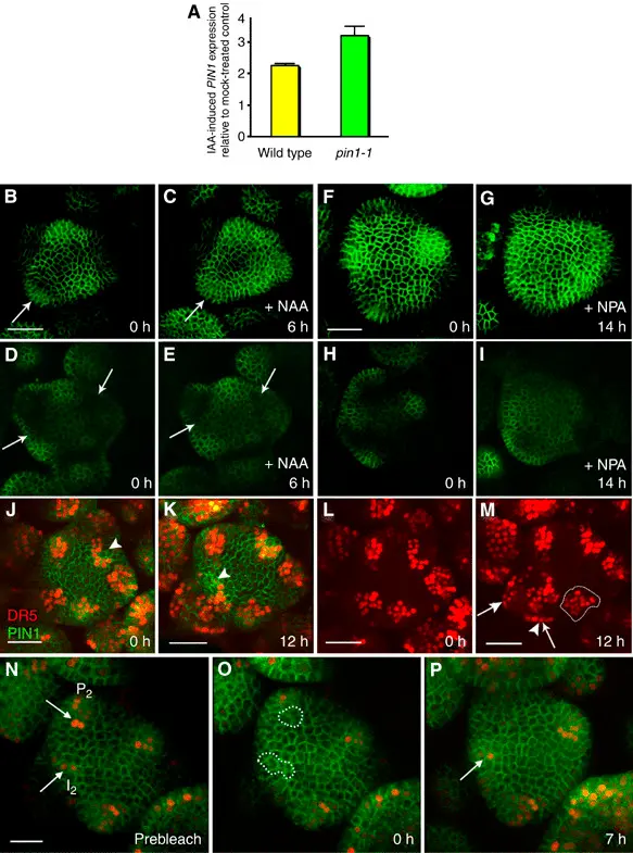
- Gene Expression Monitoring: GFP attached to promoters shows when and where genes are active. Read more
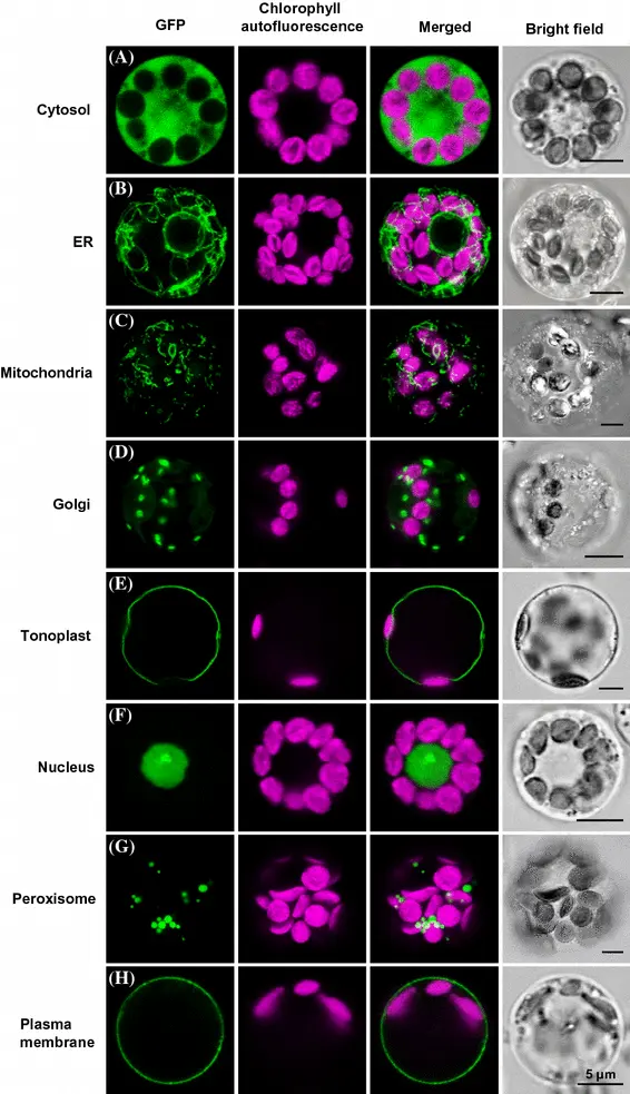
- Protein Localization: Track the movement and location of proteins inside cells. Read more
- CRISPR/Cas9 Marker: Used as a reporter to confirm successful gene editing.
- Drug Discovery: Evaluate how drugs affect live cells in real time.
GFP Variants Expand the Toolbox
Modern biotechnology has developed multiple GFP variants, including:
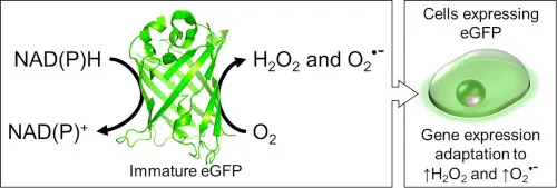
- eGFP: Enhanced brightness and stability
- CFP and YFP: Ideal for FRET experiments Read more
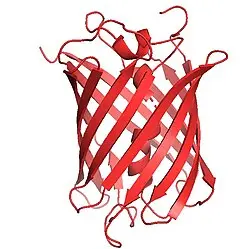
- mCherry : Enable red-shifted imaging for dual/triple labeling Read more
These fluorescent proteins allow researchers to perform multi-color imaging, making it possible to monitor several biological processes at once.
GFP in CRISPR and Gene Editing
In the era of CRISPR/Cas9 gene editing, GFP is often used as a fluorescent marker to:
- Confirm gene integration (GFP knock-in)
- Sort modified cells using flow cytometry (FACS)
- Study gene silencing and promoter activation
This ability to visualize genome editing in real time has accelerated the development of new therapies and genetic models.
Live-cell imaging using GFP enables scientists to observe:
- How cancer cells divide and spread
- How neurons form connections
- How viruses infect and replicate
Advanced microscopy techniques, such as confocal and fluorescence microscopy, rely heavily on GFP and its variants to visualize dynamic processes inside living cells.
While GFP is powerful, it comes with limitations:
- Photobleaching: Prolonged exposure to light can reduce its fluorescence.
- pH sensitivity: GFP can lose brightness in acidic environments.
However, newer variants like eGFP and mCherry have improved photostability, ensuring better results during long-term imaging.
Conclusion: The Future of GFP Biotechnology
From basic biology to high-throughput drug screening, GFP continues to illuminate the future of science. As researchers push the boundaries of genetic engineering and personalized medicine, GFP remains a key ally in making the invisible visible.
At GFPBio, we are dedicated to sharing the latest insights, tools, and applications of GFP in modern biotechnology.


