Molecular biology focuses on the fundamental processes that govern life from DNA replication and RNA transcription to protein synthesis and interaction. Yet, observing these processes in living cells remained a challenge for decades.
That changed with the discovery of Green Fluorescent Protein (GFP), a naturally fluorescent molecule derived from the jellyfish Aequorea victoria. When expressed inside cells, GFP emits a bright green glow under ultraviolet or blue light, making invisible molecular events visible in real time.
This blog explores the transformative role of GFP in molecular biology, showcasing how it has become an essential tool in gene expression analysis, protein localization, and advanced imaging techniques.
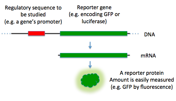
One of the most powerful uses of GFP in molecular biology is its role in tracking gene expression. By fusing the GFP gene to a promoter or gene of interest, researchers can visualize when and where that gene is activated inside a living cell.
This method allows scientists to:
- Identify tissue-specific gene expression
- Study gene regulation during development
- Monitor gene silencing or activation due to epigenetic changes
For example, in developmental biology, scientists can attach GFP to early-stage embryo genes to see when different tissues start expressing specific genes — all in real time, without harming the organism.
In molecular biology, knowing where a protein is inside the cell is just as important as knowing its structure. Proteins can travel between compartments like the nucleus, cytoplasm, or mitochondria and their location often determines their function.
GFP helps scientists track this localization by creating GFP-tagged fusion proteins. When a GFP gene is fused to the coding sequence of a target protein, it produces a fluorescent version of the protein.
This technique helps:
- Determine if a protein is nuclear, membrane-bound, or cytoplasmic
- Study protein trafficking in response to stimuli
- Observe real-time changes in protein distribution during disease
GFP has been widely used in cancer research to study the movement of mutated proteins in tumor cells.
Using GFP to Detect Protein Localization
Reporter genes are widely used in molecular biology to confirm whether a gene has been successfully expressed or edited. GFP is one of the most reliable and visible reporter genes available. Read more
In cloning, gene therapy, or CRISPR/Cas9 experiments, GFP is inserted alongside or in place of a target gene. If the modified DNA is successfully integrated and expressed, the cell glows green providing instant visual confirmation.
Benefits of using GFP as a reporter:
- Reduces the need for complex biochemical assays
- Speeds up plasmid and vector validation
- Enhances success rates in genetic engineering
Researchers can visually identify transfected cells without additional staining.
GFP has opened up a new dimension in live-cell imaging. Before GFP, most cellular observations required fixing (killing) cells. Today, scientists can use GFP to watch living cells over time using fluorescence or confocal microscopy.
This allows real-time observation of:
- Cell division (mitosis and meiosis)
- Apoptosis (programmed cell death)
- Cell migration and differentiation
GFP’s stability and low toxicity make it ideal for long-term imaging without harming cells.
Live imaging is now a staple in research fields like immunology, neuroscience, virology, and regenerative medicine.
Understanding how proteins interact is a core goal in molecular biology. GFP has been adapted into advanced techniques like FRET (Förster Resonance Energy Transfer) and Split-GFP to detect molecular interactions in living cells.
- FRET with GFP variants: Scientists attach different fluorescent proteins to two proteins of interest. If the proteins interact closely, energy transfer between the fluorophores occurs confirming interaction.
- Split-GFP: The GFP protein is split into two fragments, each fused to a different protein. Fluorescence only appears when both fragments come together due to protein interaction.
These tools are essential in:
- Signal transduction research
- Receptor-ligand studies
- Drug-target validation
Beyond research, GFP is now widely used as a teaching tool in schools and universities to help students visualize molecular biology concepts.
In classroom labs, students can:
- Use GFP-expressing bacteria to understand transformation
- Observe fluorescent gene expression under UV light
- Track plasmid uptake during genetic engineering experiments
This hands-on approach helps bridge theory and practice, making abstract concepts like transcription and translation tangible.
Conclusion: Why GFP Remains a Molecular Biology Powerhouse
Green Fluorescent Protein has done more than light up cells it has illuminated the inner workings of molecular biology. From observing gene expression to decoding protein interactions, GFP is now a foundational tool in biological research.
As new variants, sensors, and applications continue to evolve, GFP’s impact will only grow stronger making it a timeless ally in molecular exploration.
🔗 To learn more about the latest GFP-based tools and services, visit GFP Biotechnology.